
Table of Contents
- What Is a Dental X-Ray and Why Is It Taken?
- What Are the Types of Dental X-Rays?
- How Is a Dental X-Ray Taken, and Does It Hurt?
- How Much Radiation Does a Dental X-Ray Involve?
- How Does Digital Radiography Reduce Radiation?
- Can Dental X-Rays Be Taken During Pregnancy and Breastfeeding?
- Are Dental X-Rays Safe for Children?
- What Protective Measures Are Taken During X-Ray Imaging?
- How Often Should Dental X-Rays Be Taken?
- Frequently Asked Questions About Dental X-Rays
What Is a Dental X-Ray and Why Is It Taken?
"Is this X-ray going to harm me?" is one of the most common concerns voiced in the dental chair. The worry is understandable, yet the imaging methods used in modern dentistry operate with extremely low radiation doses and are only applied when genuinely necessary for diagnosis or treatment.
A dental X-ray makes visible what a visual examination cannot see. Cavities starting between the teeth, infections at the root tips, bone loss in the jaw, cysts and similar lesions, and teeth that have not yet erupted can only be reliably assessed with radiographic imaging. For example, the position of wisdom teeth in the jaw and their effect on neighbouring teeth cannot be determined dependably without an X-ray.
An X-ray is therefore not an "extra procedure" but one of the cornerstones of an accurate diagnosis and a sound treatment plan. Treatment carried out without imaging risks missing the true source of the problem. X-ray records also make it possible to follow the success of treatment objectively: whether bone healing has taken place or an infection has regressed can often only be understood by comparison with earlier images.
What Are the Types of Dental X-Rays?
There is no single type of dental X-ray; each imaging method has a different purpose and scope. The dentist chooses the technique that provides the most information with the least exposure for the clinical situation:
- Periapical X-ray: Shows one or a few teeth in detail together with their roots; used to assess root-tip infections and deep decay.
- Bitewing X-ray: Shows the crowns of the upper and lower teeth in a single frame; preferred for the early detection of cavities between the teeth.
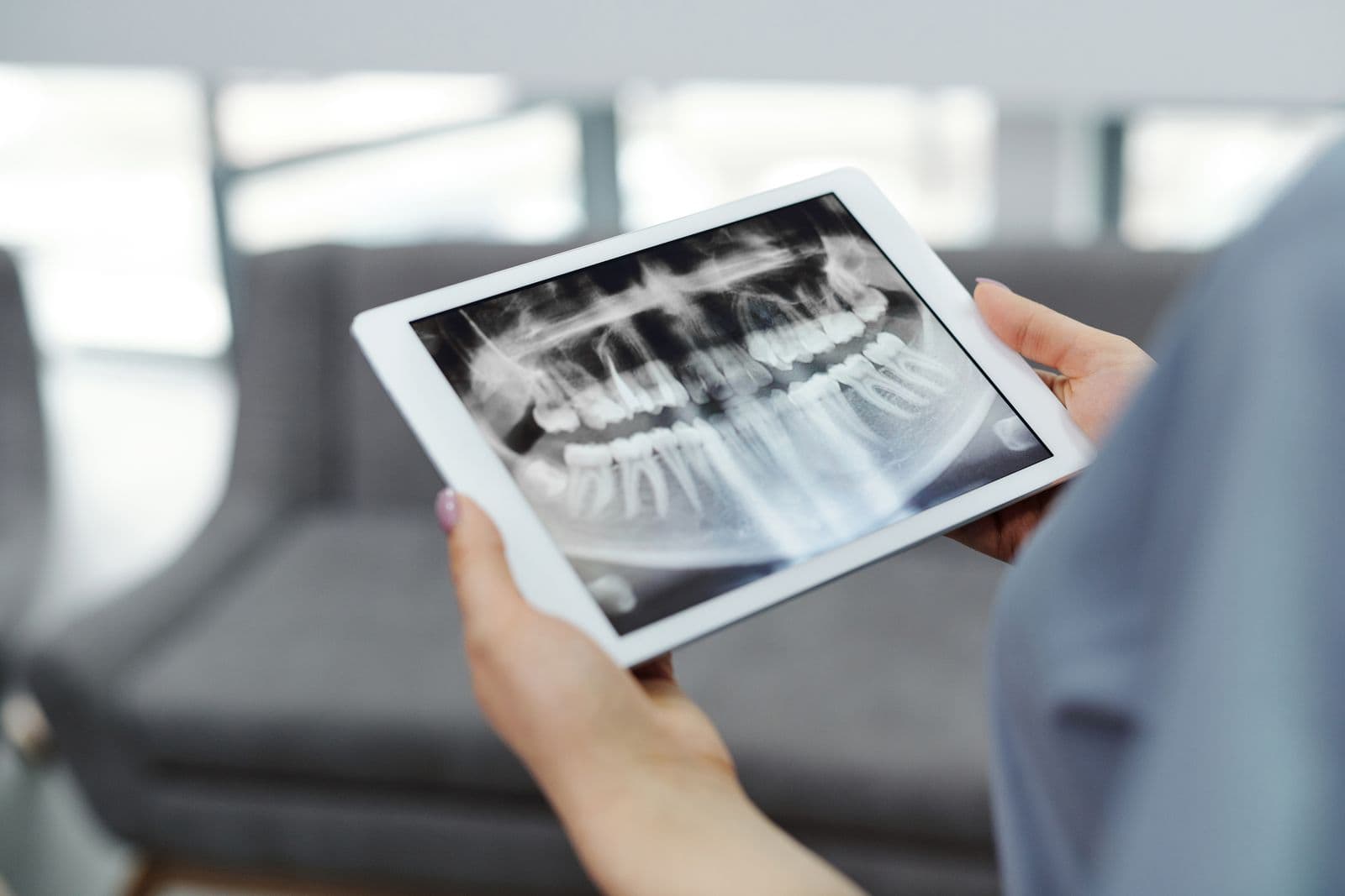
- Panoramic X-ray: Presents all the teeth, the jawbones and the jaw joints in one image; widely used for general assessment, impacted teeth and treatment planning.
- Cephalometric X-ray: Provides a side view of the head and jaw structure; mostly used in orthodontic treatment planning.
- Cone-beam computed tomography (CBCT): Creates a three-dimensional image of the jaws and teeth; provides detailed information for implant planning, impacted-tooth surgery and complex cases.
How Is a Dental X-Ray Taken, and Does It Hurt?
A dental X-ray is one of the fastest and least demanding procedures in medical imaging. For intraoral images, a small digital sensor or film is placed on the inner side of the tooth to be imaged, and the exposure is completed within a few seconds. The sensor stays in the mouth only briefly; although a mild gag reflex may be triggered in some people, the procedure is painless and involves no needles, incisions or any similar intervention.
For a panoramic image, nothing is placed in the patient's mouth at all: the patient stands or sits still in front of the device, the arm of the machine rotates slowly around the head, and the exposure is completed in about 15-20 seconds. Before the scan, you will be asked to remove items that could affect the image, such as metal jewellery, glasses and hearing aids. CBCT imaging is likewise painless and usually takes less than a minute.
In short, contrary to the perception of a "difficult procedure" in many patients' minds, an X-ray is a natural and comfortable part of the examination; all you need to do during the exposure is stay still for a few seconds.
How Much Radiation Does a Dental X-Ray Involve?
Radiation doses are measured in millisieverts (mSv), and dental X-rays are among the lowest-dose procedures in medical imaging. The literature reports the dose of a single digital periapical or bitewing X-ray at approximately 0.005 mSv, and that of a panoramic X-ray at roughly 0.01-0.02 mSv.
Comparing these figures with daily life puts them in perspective: the human body is continuously exposed to natural background radiation from soil, building materials, food and cosmic rays, and this natural exposure averages about 0.008 mSv per day. A single small dental X-ray therefore corresponds to roughly one day of natural background radiation, or an exposure close to that received during a flight of a few hours. For comparison, the dose of a single chest X-ray is around 0.1 mSv, so a panoramic dental X-ray involves less than a tenth of that exposure. In the assessments of international radiation-safety bodies, exposures at this level are not associated with any measurable health risk.
The dose of a three-dimensional CBCT scan varies with the size of the imaged area but is higher than that of conventional X-rays. For this reason, CBCT is not used as a routine screening tool but as an additional source of information when two-dimensional images are insufficient.
How Does Digital Radiography Reduce Radiation?
Digital sensors, which have replaced old film-based X-rays, need far less exposure to form an image; digital systems are reported to reduce the dose markedly compared with film, by more than half according to many sources.
The advantage of digital imaging is not only the low dose. The image appears on screen within seconds, fine details can be examined by magnifying the image and adjusting the contrast, and the need for repeat exposures decreases. Because the images are archived, changes over time can also be followed comparatively. You can read in detail how these possibilities translate into treatment in our article on digital dentistry in practice.
Can Dental X-Rays Be Taken During Pregnancy and Breastfeeding?
Pregnancy is the period in which concerns about X-rays are felt most strongly. International obstetric and dental organisations accept that dental X-rays can be taken during pregnancy when the necessary protective measures are in place; the imaged area is far from the abdomen, and a lead apron provides additional protection.
In practice, however, the guiding principle is this: non-urgent exposures that can wait are generally postponed until after the birth or to a more suitable stage of the pregnancy, while imaging is not delayed in conditions such as pain, infection or abscess that threaten the mother's health, because the risk posed by an untreated infection to mother and baby is far greater than the hypothetical risk of a low-dose X-ray. You can find our full care recommendations for this period in our dental health during pregnancy guide.
During breastfeeding the situation is even clearer: the rays used in dental radiography do not accumulate in the body and do not affect breast milk, so there is no known objection to dental X-rays in breastfeeding mothers.
Are Dental X-Rays Safe for Children?
In children, the decision to take an X-ray is made with even greater care than in adults. Because developing tissues are more sensitive to radiation, imaging in children is performed only when there is a question the clinical examination cannot answer; it is not applied as a routine habit.
At the same time, the information an X-ray provides in children is often critical: hidden cavities between primary teeth, eruption problems, the condition of roots after trauma and the development of the permanent tooth germs can only be assessed with imaging. When exposures are made with child-specific dose settings, short exposure times and protective aprons, the diagnostic benefit clearly outweighs the extremely low risk. How often images are needed is planned individually by the dentist according to the child's caries risk and stage of development. To keep the process from being frightening for the child, the procedure is explained in advance; in young children who find it hard to cooperate, imaging can be postponed to a time the child can adapt to, provided there is no urgency.
What Protective Measures Are Taken During X-Ray Imaging?
In modern clinics, radiation safety rests on the internationally accepted ALARA principle: "as low as reasonably achievable". The main measures applied under this principle are:
- Justification principle: An X-ray is taken only if it will provide information that changes the diagnosis or treatment; no images are taken "just in case".
- Lead apron and thyroid collar: They cover radiation-sensitive organs and reduce exposure outside the imaged area.
- Choosing the right technique: The lowest-dose method that images the smallest sufficient area is preferred for the problem at hand.
- Digital systems and collimation: Focusing the beam strictly on the relevant region and using sensitive sensors keeps the dose to a minimum.
- Equipment calibration: X-ray devices undergo regular maintenance and quality-control checks. In addition, the training of the team taking the images and correct patient positioning reduce the need for repeat exposures and thus lower the total dose.
How Often Should Dental X-Rays Be Taken?
There is no single answer that applies to everyone; the frequency of imaging is determined individually according to oral health status, caries risk and the treatment being carried out. In an adult with good oral health and low caries risk, check-up X-rays at long intervals are generally sufficient, while closer follow-up may be needed in people with active decay or gum disease. Similarly, in people undergoing orthodontic treatment, in the follow-up of gum disease and in the monitoring of teeth that have suffered trauma, control images may form part of the treatment plan. Rather than automatically requesting a new image at every visit, your dentist compares your previous images and decides whether a new exposure is genuinely necessary.
Some treatments require imaging by their very nature: the three-dimensional assessment of bone volume and anatomical structures before implant treatment, and checking the length of the root canals and the accuracy of the filling during root canal treatment, are impossible without X-rays. In these situations imaging is part of the safety of the treatment itself, and its benefit far exceeds the dose involved.
Frequently Asked Questions About Dental X-Rays
Do dental X-rays cause cancer? Scientific data do not show that dental X-rays taken for diagnostic purposes and when needed lead to a measurable increase in cancer risk. The doses are close to daily levels of natural background radiation; nevertheless, as with any exposure, the justification principle is followed and unnecessary imaging is avoided.
What is the difference between a panoramic X-ray and CBCT? A panoramic X-ray provides a two-dimensional overview of the whole mouth at a low dose, while CBCT offers three-dimensional, cross-sectional images at a higher dose. Which method is needed is assessed through a clinical examination, depending on the nature of the problem.
I had a dental X-ray while pregnant - will it harm my baby? The area exposed during a dental X-ray is far from the abdomen and the doses are very low; there is no scientific evidence that a single exposure taken with a lead apron harms the baby. Even so, discussing your concerns with your obstetrician will give you reassurance.
Why do I wear a lead apron during a dental X-ray? Lead is a material that X-rays cannot pass through; the apron and thyroid collar are used to minimise the exposure of radiation-sensitive organs outside the imaged area. This does not mean the dose is dangerous; it is a requirement of the precautionary approach.
Is it harmful to have several X-rays within a short time? During treatment, for example at different stages of a root canal, several small X-rays may be necessary, and their combined dose still remains low. Your dentist plans every exposure on the basis of diagnostic need; your individual situation is assessed through a clinical examination.
Can I have a dental X-ray while breastfeeding? Yes; the X-rays used in dental imaging do not accumulate in the body or in breast milk. There is no known objection to dental radiography during breastfeeding; your treatment plan is tailored to you through a clinical examination.

This content was prepared by the dental team of our clinic in Cukurambar, Ankara. Feel free to contact us with any questions about your oral and dental health.
This content is for informational purposes only; a dental examination is required for diagnosis and treatment.
Have questions? We are happy to help.
Related Articles

Why Do Teeth Turn Yellow? Causes of Tooth Discoloration and Solutions
Is it tea, coffee and smoking, or age, medication and enamel structure? The extrinsic and intrinsic causes of yellow teeth, the limits of home remedies, professional solutions and the habits that slow discoloration, all in this guide.
Gum Inflammation (Gingivitis): Symptoms, Causes and Treatment
Gums that bleed while brushing and look red and swollen are often the first sign of gingivitis. What causes gum inflammation, how does it differ from periodontitis, and how is it treated? Symptoms, treatment and home care in this guide.
Dental Bonding: What Is Composite Bonding and How Is It Done?
A chipped edge, a small gap or a stain that whitening cannot remove can often be fixed in a single visit. The steps of dental bonding, its longevity, how it differs from veneers and aftercare tips — all in this guide.