
Table of Contents
- Why Do Wisdom Teeth Cause Problems?
- What Are Impacted and Partially Impacted Teeth?
- Pericoronitis: The Most Common Complaint
- When Is Extraction Recommended — and When Is Monitoring Enough?
- Watchful Waiting: Not Every Impacted Tooth Comes Out
- The Role of X-Rays: Imaging Sharpens the Decision
- How Does Extraction or Impacted Tooth Surgery Proceed?
- Recovery: What to Watch For, Including Dry Socket
- Effects on Neighbouring Teeth — and the Bottom Line
If your wisdom tooth has started to ache, you are probably asking one question: does it have to come out, or can it stay? The short answer is that not every wisdom tooth needs extraction. The decision depends on the tooth's position, your jaw anatomy, your oral hygiene and the X-ray findings — and it is made individually by your dentist.
Wisdom teeth, or third molars, are the last permanent teeth to erupt, usually between the ages of 17 and 25. Because the modern human jaw often lacks space for them, eruption problems are extremely common. Common, however, does not mean that surgery is always required.
In this article we walk through why wisdom teeth cause trouble, what an impacted tooth actually is, which criteria guide the extraction decision, how the procedure works, and what to watch for during recovery.
Why Do Wisdom Teeth Cause Problems?
The root of the problem is lack of space. Over the course of evolution our jaws became smaller while the number of teeth stayed the same, so the last teeth to arrive often struggle to find room. A tooth without space may remain inside the jawbone, lean at an angle against the neighbouring molar, or break only partially through the gum.
A partially erupted or tilted wisdom tooth creates an area that is very difficult to clean with a toothbrush or floss. Plaque accumulating there can pave the way for decay, gum inflammation and bad breath. And the damage may not stop at the wisdom tooth itself — the second molar in front of it can be affected as well.
On the other hand, in people with sufficient jaw space, wisdom teeth can erupt completely normally, take part in chewing and remain trouble-free for life. The blanket statement that wisdom teeth always have to be removed is simply not scientifically accurate.
What Are Impacted and Partially Impacted Teeth?
An impacted tooth is one that has failed to emerge into the mouth even though its normal eruption time has passed, remaining trapped in the jawbone or under the gum. If the tooth lies entirely within bone it is called fully impacted; if part of it has broken through the gum it is partially impacted (or partially erupted).
Impacted teeth are also classified by position: vertical (correctly oriented but unerupted), mesioangular (tilted forward against the neighbouring tooth), horizontal (lying completely sideways) and distoangular (tilted backward) are the most common patterns. The angle and depth of the tooth influence both the likelihood of problems and the complexity of any surgery.
Clinically, partially impacted teeth tend to be the riskiest group. They are exposed to oral bacteria because they communicate with the mouth, yet they cannot be cleaned properly because they never fully erupted. That combination is one of the most frequent causes of recurring inflammation.
Pericoronitis: The Most Common Complaint
Pericoronitis is an inflammation that develops when bacteria and food debris collect under the flap of gum tissue (the operculum) partially covering a semi-erupted tooth. Typical signs include pain, swelling, redness, difficulty opening the mouth and discomfort when swallowing; in some cases a mild fever may accompany the picture.
In the acute phase, your dentist will usually aim to calm the inflammation by cleaning the area, prescribing antiseptic rinses and, where appropriate, medication. Pericoronitis, however, has a strong tendency to recur: as long as the tooth remains partially impacted, the same episode can repeat itself. Recurrent pericoronitis is therefore one of the key findings that supports an extraction decision.
If you notice symptoms of pericoronitis, it is important to see a dentist rather than trying to treat the area yourself — only a clinical examination can assess whether the infection may spread to surrounding tissues.
When Is Extraction Recommended — and When Is Monitoring Enough?
The decision to extract is never based on a single criterion; it rests on the combined assessment of the clinical examination and radiographic findings. The evidence-based approach is to remove teeth that are causing problems or are highly likely to, and to monitor trouble-free teeth at regular intervals.
Extraction is typically considered in situations such as:
- Recurrent episodes of pericoronitis
- Decay in the wisdom tooth or the adjacent second molar in a position that cannot be restored
- Pressure on the neighbouring root with signs of root resorption
- Suspicion of a cyst or other pathological lesion around the tooth
- Space requirements within an orthodontic treatment plan
- A position that interferes with denture or implant planning
- A partially erupted position that cannot be kept clean and keeps getting inflamed
Watchful Waiting: Not Every Impacted Tooth Comes Out
For a fully impacted wisdom tooth that sits symmetrically within the bone, shows no surrounding pathology and does not harm the neighbouring tooth, regular monitoring can often be sufficient. The idea that every symptom-free impacted tooth will inevitably cause trouble later is a myth; current guidance favours individual risk assessment over routine prophylactic removal of asymptomatic, disease-free teeth.
A monitoring decision does not mean the tooth is forgotten. At the intervals your dentist recommends — usually annual check-ups with X-rays when needed — the tooth's position, the surrounding bone and the health of the adjacent molar are reviewed. If the picture changes, the decision can be revisited.
The essential point is this: the choice between extraction and monitoring is not made from general information on the internet, but by the dentist who has examined your mouth and evaluated your own X-rays.
The Role of X-Rays: Imaging Sharpens the Decision
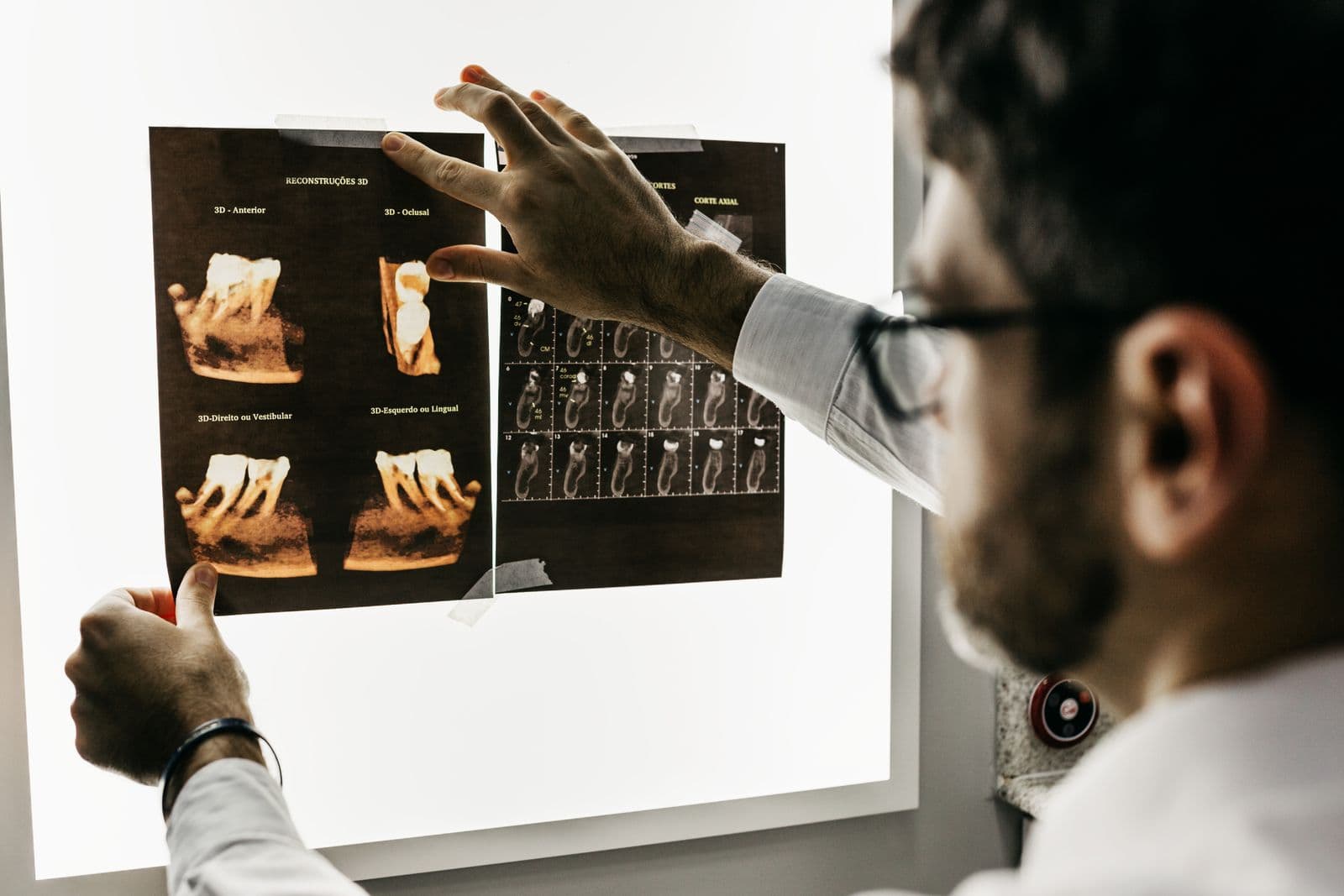
A panoramic X-ray is the standard starting point for assessing wisdom teeth. In a single image it shows the tooth's angle and depth, the number and shape of its roots, its relationship to the neighbouring molar and any pathology in the surrounding bone.
For lower wisdom teeth whose roots lie close to the mandibular nerve — the nerve that supplies sensation to the lower lip and chin — three-dimensional imaging (cone-beam computed tomography, CBCT) may be requested. CBCT shows the true distance between the root and the nerve canal in millimetres, allowing the surgical plan to be tailored accordingly. In the upper jaw, the relationship between the tooth and the sinus floor is evaluated in a similar way.
In short, imaging answers both questions: whether the tooth should be removed or monitored, and — if removal is chosen — how it should be done safely.
How Does Extraction or Impacted Tooth Surgery Proceed?
Removing a normally erupted wisdom tooth is usually no different from a routine extraction and can be completed quickly under local anaesthesia. Surgery for an impacted tooth is a minor surgical procedure: the area is fully numbed with local anaesthetic, a small incision is made in the gum, a thin layer of bone over the tooth is removed if necessary, and the tooth is sometimes divided and taken out in sections. The site is then irrigated and closed with sutures.
Thanks to the anaesthesia you will not feel pain during the procedure; sensations of pressure and vibration are normal. The duration depends on the tooth's position and depth — many cases are completed within 20 to 45 minutes. For patients with significant anxiety, sedation options can be discussed if the dentist considers them appropriate.
Before the procedure, make sure to tell your dentist about any medications you take, chronic conditions and allergies, as the surgical plan is shaped around this information. At clinics such as ADEN Dental in Çukurambar, Ankara, this assessment can often be planned together with the examination and imaging in a single visit.
Recovery: What to Watch For, Including Dry Socket
The first 24 to 48 hours after extraction are the most critical phase of healing. The blood clot that forms in the socket is the wound's natural dressing; protecting it both speeds healing and is the key to preventing alveolar osteitis, better known as dry socket. Dry socket develops when the clot is lost early and the bone is left exposed; it typically announces itself two to four days after the extraction with intensifying pain that may radiate towards the ear — a situation in which you should contact your dentist.
To make recovery as smooth as possible, you can follow these recommendations:
- Bite firmly on the gauze pad for the first 30-45 minutes without interruption, then remove it
- For the first 24 hours do not spit, rinse or use a straw
- Avoid smoking and all tobacco products for at least 72 hours — they markedly increase the risk of dry socket
- Apply cold packs externally on the first day at intervals (15 minutes on, then a break)
- Choose lukewarm, soft foods in the first days and avoid very hot food and drinks
- Take any medication your dentist prescribes exactly as directed
- From the next day on, keep brushing gently while avoiding the extraction site
Effects on Neighbouring Teeth — and the Bottom Line
A tilted wisdom tooth can create an uncleanable niche against the back surface of the second molar and contribute to decay there; more rarely, it can press on the neighbouring root and cause resorption. The popular belief that wisdom teeth push the front teeth and cause crowding is less clear-cut than it seems: research shows that lower front crowding has many contributing factors and cannot be attributed to wisdom teeth alone.
In summary, wisdom teeth are neither problem teeth that must automatically be removed nor structures that can simply be ignored. The right approach is an individual assessment with examination and X-rays, a planned extraction when it is needed, and regular monitoring when it is not. If you are experiencing pain, swelling or eruption problems with a wisdom tooth — or simply want to know where you stand — a dental examination will give you a clear, personalised roadmap.

This content was prepared by the dental team of our clinic in Cukurambar, Ankara. Feel free to contact us with any questions about your oral and dental health.
This content is for informational purposes only; a dental examination is required for diagnosis and treatment.
Have questions? We are happy to help.
Related Articles
Orthodontic Treatment in Children: At What Age Are Braces Fitted?
Have you noticed crowding in your child's teeth? The right time for the first orthodontic check-up, the causes of crowding, the benefits of early treatment, appliance types and braces care — all in this comprehensive family guide.
What Is a Dental Bridge? Types, Procedure Steps and Comparison with Implants
Considering a bridge or an implant for your missing teeth? In this comprehensive guide you will find the types of dental bridges, the step-by-step procedure, the finer points of cleaning under a bridge and a comparison with implants.
Are Dental X-Rays Harmful? A Guide to Radiation Doses and Safety
Worried about getting a dental X-ray? This guide covers the actual radiation doses of panoramic X-rays and CBCT scans, safety principles during pregnancy and childhood, and the protective measures taken in modern clinics.